Now Reading: Understanding Phyllodes Tumor: Diagnosis and Treatment
-
01
Understanding Phyllodes Tumor: Diagnosis and Treatment
Understanding Phyllodes Tumor: Diagnosis and Treatment
Phyllodes tumors, also known as cystosarcoma phyllodes, are rare fibroepithelial neoplasms that primarily arise in the breast. These tumors are characterized by their leaf-like architecture, which is where the name “phyllodes” originates from the Greek word for “leaf.” Although they can occur in other tissues, such as the skin and soft tissues, the breast is the most common site of occurrence. Phyllodes tumors are classified as benign, malignant, or borderline based on their histological features and behavior.
The incidence of phyllodes tumors is relatively low, accounting for approximately 0.3% to 1% of all breast tumors. They typically affect women in their 30s to 50s, although they can occur at any age. The growth rate of these tumors can be rapid, often leading to significant size before diagnosis.
Due to their unique characteristics and potential for aggressive behavior, understanding phyllodes tumors is crucial for effective management and treatment.
Key Takeaways
- Phyllodes tumor is a rare type of breast tumor that forms in the connective tissue of the breast.
- Symptoms of Phyllodes tumor may include a painless breast lump, breast pain, and changes in breast size or shape.
- Diagnosis of Phyllodes tumor involves a physical exam, imaging tests, and a biopsy to confirm the presence of the tumor.
- Imaging tests such as mammogram, ultrasound, and MRI are used to determine the size and location of the tumor.
- Treatment options for Phyllodes tumor may include surgery, radiation therapy, and follow-up care for monitoring and prognosis.
Symptoms and Risk Factors
Patients with phyllodes tumors often present with a palpable mass in the breast, which may be painless or associated with discomfort. The tumor can vary in size, ranging from a few centimeters to over 10 centimeters in diameter. Other symptoms may include changes in breast shape or contour, skin dimpling, or nipple discharge.
In some cases, patients may experience lymphadenopathy if the tumor has metastasized to regional lymph nodes. Several risk factors have been identified that may predispose individuals to develop phyllodes tumors. These include a family history of breast cancer, previous radiation exposure to the chest area, and certain genetic syndromes such as Li-Fraumeni syndrome or Cowden syndrome.
Additionally, hormonal factors may play a role, as some studies suggest that estrogen exposure could influence tumor development. However, the exact etiology remains largely unclear, necessitating further research into the underlying mechanisms.
Diagnosis of Phyllodes Tumor
The diagnosis of phyllodes tumors typically begins with a thorough clinical examination and imaging studies. A detailed medical history is essential to assess any risk factors or previous breast conditions. Physical examination often reveals a firm, mobile mass that may be well-defined or irregular in shape.
Imaging modalities such as mammography and ultrasound are commonly employed to evaluate breast masses. Mammography may show a well-circumscribed mass with lobulated contours, while ultrasound can provide additional information regarding the tumor’s internal characteristics. However, definitive diagnosis often requires histopathological evaluation through biopsy.
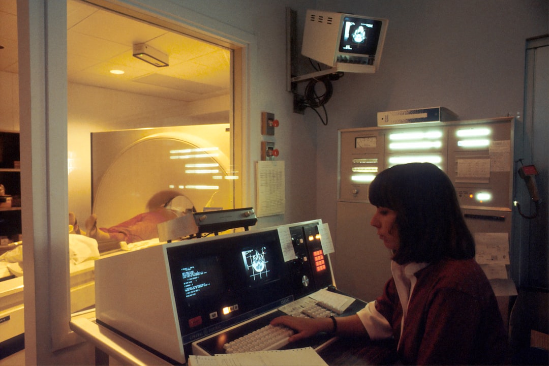
Imaging Tests for Phyllodes Tumor
| Imaging Test | Accuracy | Advantages | Disadvantages |
|---|---|---|---|
| Ultrasound | Variable | Non-invasive, widely available | Operator-dependent, limited in larger tumors |
| MRI | High | Excellent soft tissue contrast | Expensive, time-consuming |
| CT Scan | Low | Good for detecting calcifications | Ionizing radiation, limited soft tissue contrast |
Imaging tests play a pivotal role in the initial assessment and ongoing management of phyllodes tumors. Mammography is typically the first-line imaging modality used to identify breast masses. In cases of phyllodes tumors, mammograms may reveal a well-defined mass with lobulated margins and possible calcifications.
However, due to the variable appearance of these tumors, mammography alone may not provide sufficient information for diagnosis. Ultrasound is another valuable tool that can help differentiate between solid and cystic masses. It provides real-time imaging and can guide needle biopsies for histological analysis.
MRI may also be utilized in certain cases to assess tumor extent and involvement of surrounding tissues, particularly when surgical planning is necessary. The combination of these imaging modalities enhances diagnostic accuracy and aids in determining the appropriate management strategy.
Biopsy and Pathology of Phyllodes Tumor
Biopsy is essential for confirming the diagnosis of phyllodes tumors and involves obtaining tissue samples for histopathological examination. There are several biopsy techniques available, including fine-needle aspiration (FNA), core needle biopsy, and excisional biopsy.
Histopathological evaluation reveals distinct features of phyllodes tumors, including stromal overgrowth and a characteristic leaf-like architecture. The presence of stromal atypia, mitotic activity, and necrosis are critical factors in determining the tumor’s grade and behavior. Pathologists classify phyllodes tumors into benign, borderline, or malignant categories based on these histological characteristics, which significantly influence treatment decisions and prognosis.
Staging and Grading of Phyllodes Tumor

Staging and grading are crucial components in the management of phyllodes tumors, as they provide insight into tumor behavior and potential outcomes. The grading system primarily focuses on histological features such as stromal cellularity, nuclear atypia, and mitotic activity. Benign tumors exhibit low cellularity and minimal atypia, while malignant tumors demonstrate high cellularity with significant atypical features.
Staging involves assessing the extent of disease spread beyond the primary tumor site. This includes evaluating regional lymph nodes and distant metastasis through imaging studies such as CT scans or PET scans. Accurate staging is essential for determining appropriate treatment options and predicting patient outcomes.
Treatment Options for Phyllodes Tumor
The primary treatment modality for phyllodes tumors is surgical excision, which aims to achieve clear margins and minimize recurrence risk. The extent of surgery depends on the tumor’s size, grade, and location. In cases of benign tumors, wide local excision may suffice; however, malignant tumors often necessitate more extensive surgical intervention.
Chemotherapy is generally not effective for phyllodes tumors due to their unique biology; however, targeted therapies may be explored in clinical trials for advanced cases.
Surgery for Phyllodes Tumor
Surgical intervention remains the cornerstone of treatment for phyllodes tumors. The goal of surgery is to remove the tumor along with a margin of healthy tissue to reduce the likelihood of local recurrence. Surgical options include lumpectomy or mastectomy depending on tumor size and location.
In cases where the tumor is large or has infiltrative characteristics, mastectomy may be necessary to ensure complete removal. Sentinel lymph node biopsy may also be performed if there is concern about lymphatic spread; however, routine axillary dissection is not typically indicated unless there is evidence of metastasis.
Radiation Therapy for Phyllodes Tumor
Radiation therapy may be utilized as an adjunct treatment for patients with malignant phyllodes tumors or those with close surgical margins after resection. The rationale behind radiation therapy is to target residual tumor cells that may remain post-surgery, thereby reducing the risk of local recurrence. Radiation treatment typically involves external beam radiation delivered over several weeks.
The dosage and schedule depend on individual patient factors and tumor characteristics. While radiation therapy has shown promise in improving local control rates, its role remains controversial due to limited data on long-term outcomes.
Follow-Up Care and Monitoring
Post-treatment follow-up care is essential for monitoring potential recurrence or metastasis of phyllodes tumors. Patients should undergo regular clinical examinations every 3 to 6 months during the first few years after treatment, followed by annual evaluations thereafter. Imaging studies may also be warranted based on clinical findings or if there are concerns regarding recurrence.
Patient education regarding self-examination techniques and awareness of any new symptoms is crucial for early detection of potential issues.
Prognosis and Outlook for Phyllodes Tumor
The prognosis for patients with phyllodes tumors varies significantly based on tumor grade and stage at diagnosis. Benign phyllodes tumors generally have an excellent prognosis with low recurrence rates following complete surgical excision. In contrast, malignant phyllodes tumors carry a higher risk of local recurrence and distant metastasis.
Overall survival rates for patients with malignant phyllodes tumors can range from 50% to 80%, depending on various factors such as tumor size, grade, and response to treatment. Continuous research into the biology of these tumors aims to improve understanding and develop more effective therapeutic strategies. In summary, phyllodes tumors are rare breast neoplasms characterized by their unique histological features and potential for aggressive behavior.
Early diagnosis through imaging and biopsy is crucial for effective management. Surgical excision remains the primary treatment modality, with radiation therapy considered in select cases. Ongoing follow-up care is essential for monitoring recurrence risk and ensuring optimal patient outcomes.
Phyllodes tumors are rare breast tumors that can be challenging to diagnose and treat. For more information on breast health and cancer awareness, check out this article on celebrity spotlight featuring stories of celebrities who have battled breast cancer. It is important to stay informed and proactive about your health to catch any potential issues early.
FAQs
What is a phyllodes tumor?
A phyllodes tumor is a rare type of breast tumor that forms in the connective tissue of the breast. It can be benign, borderline, or malignant.
What are the symptoms of a phyllodes tumor?
Symptoms of a phyllodes tumor may include a painless lump in the breast, breast pain, and changes in breast size or shape.
How is a phyllodes tumor diagnosed?
Diagnosis of a phyllodes tumor typically involves a physical examination, imaging tests such as mammograms or ultrasounds, and a biopsy to examine the tissue under a microscope.
What are the treatment options for a phyllodes tumor?
Treatment for a phyllodes tumor may involve surgery to remove the tumor, and in some cases, radiation therapy or chemotherapy may be recommended.
What is the prognosis for a phyllodes tumor?
The prognosis for a phyllodes tumor depends on various factors including the size of the tumor, whether it is benign, borderline, or malignant, and how early it is diagnosed and treated.
Are phyllodes tumors common?
Phyllodes tumors are rare, accounting for less than 1% of all breast tumors. They most commonly occur in women in their 40s, but can also occur in younger women and men.