Now Reading: Understanding Glioma Brain Tumors: Symptoms, Diagnosis, and Treatment
-
01
Understanding Glioma Brain Tumors: Symptoms, Diagnosis, and Treatment
Understanding Glioma Brain Tumors: Symptoms, Diagnosis, and Treatment
A glioma brain tumor is a type of neoplasm that originates from glial cells, which are supportive cells in the central nervous system (CNS). These tumors can arise in various regions of the brain and spinal cord, and they are classified based on the specific type of glial cell from which they develop. The primary glial cell types include astrocytes, oligodendrocytes, and ependymal cells.
Gliomas account for approximately 30% of all brain tumors and about 80% of all malignant brain tumors, making them a significant concern in neuro-oncology. The World Health Organization (WHO) classifies gliomas into four grades based on their histological characteristics and aggressiveness. Grade I tumors are benign and slow-growing, while Grade IV tumors, such as glioblastomas, are highly malignant and aggressive.
The prognosis for patients diagnosed with gliomas varies significantly depending on the tumor’s grade, location, and the patient’s overall health. Early detection and intervention are crucial for improving outcomes.
Key Takeaways
- Glioma brain tumors are a type of tumor that originates in the brain and spinal cord.
- There are different types of glioma brain tumors, including astrocytomas, oligodendrogliomas, and ependymomas.
- Symptoms of glioma brain tumors can include headaches, seizures, and changes in cognitive function.
- Risk factors for glioma brain tumors may include exposure to radiation and certain genetic conditions.
- Diagnosing glioma brain tumors involves a combination of imaging tests, biopsies, and pathology evaluations.
Types of Glioma Brain Tumors
Gliomas can be categorized into several distinct types, each with unique characteristics and clinical implications. The most common types include: 1. **Astrocytomas**: These tumors arise from astrocytes and can range from low-grade (Grade I and II) to high-grade (Grade III and IV).
Glioblastoma multiforme (GBM), a Grade IV astrocytoma, is the most aggressive form and is associated with a poor prognosis. 2. **Oligodendrogliomas**: Originating from oligodendrocytes, these tumors typically present in the cerebral hemispheres.
They are often classified as low-grade (Grade II) or anaplastic (Grade III), with the latter being more aggressive. 3. **Ependymomas**: These tumors develop from ependymal cells lining the ventricles of the brain and the spinal canal.
They can occur in both children and adults and are classified into various grades based on their histological features. 4. **Mixed Gliomas**: These tumors contain a combination of different glial cell types, such as astrocytes and oligodendrocytes.
An example is an anaplastic oligoastrocytoma, which exhibits characteristics of both astrocytomas and oligodendrogliomas. Understanding the specific type of glioma is essential for determining the appropriate treatment strategy and predicting patient outcomes.
Symptoms of Glioma Brain Tumors
The clinical presentation of glioma brain tumors can vary widely depending on the tumor’s size, location, and growth rate. Common symptoms include: 1. **Headaches**: Persistent or worsening headaches are often reported by patients with gliomas.
These headaches may be accompanied by nausea or vomiting due to increased intracranial pressure. 2. **Neurological Deficits**: Depending on the tumor’s location, patients may experience focal neurological deficits such as weakness, sensory loss, or difficulty with coordination and balance.
3. **Cognitive Changes**: Gliomas can affect cognitive function, leading to memory problems, confusion, or changes in personality. Patients may also experience difficulties with speech or language.
4. **Seizures**: Seizures are a common symptom associated with gliomas, particularly in cases where the tumor irritates surrounding brain tissue. 5. **Visual Disturbances**: Tumors located near the optic pathways may cause visual disturbances, including blurred vision or loss of vision. Recognizing these symptoms early can facilitate timely diagnosis and treatment, ultimately improving patient outcomes.
Risk Factors for Glioma Brain Tumors
| Risk Factor | Description |
|---|---|
| Age | Glioma tumors are more common in older adults. |
| Genetics | Family history of glioma or certain genetic conditions can increase the risk. |
| Exposure to radiation | Previous radiation treatment to the head may increase the risk. |
| Chemical exposure | Exposure to certain chemicals or toxins may be a risk factor. |
| Cell phone use | Some studies suggest a possible link between long-term cell phone use and glioma risk. |
While the exact etiology of gliomas remains largely unknown, several risk factors have been identified that may increase an individual’s likelihood of developing these tumors: 1. **Genetic Predisposition**: Certain genetic syndromes, such as neurofibromatosis type 1 (NF1) and Li-Fraumeni syndrome, are associated with an increased risk of gliomas. Additionally, mutations in specific genes like TP53 and IDH1 have been linked to glioma development.
2. **Age**: Gliomas can occur at any age; however, they are more prevalent in adults between the ages of 45 and 70 years. Pediatric gliomas often present differently than those in adults.
3. **Exposure to Ionizing Radiation**: Individuals who have undergone radiation therapy for other cancers, particularly to the head or neck region, have a higher risk of developing secondary gliomas. 4.
**Environmental Factors**: Some studies suggest that exposure to certain chemicals or pesticides may be associated with an increased risk of glioma; however, further research is needed to establish definitive links. 5. **Gender**: Epidemiological studies indicate that males are more likely to develop gliomas than females, although the reasons for this disparity remain unclear.
Awareness of these risk factors can aid in identifying individuals who may benefit from closer monitoring or preventive strategies.
Diagnosing Glioma Brain Tumors
The diagnostic process for glioma brain tumors typically begins with a thorough medical history and neurological examination. Physicians assess symptoms and perform cognitive tests to evaluate brain function. If glioma is suspected, several diagnostic modalities may be employed: 1.
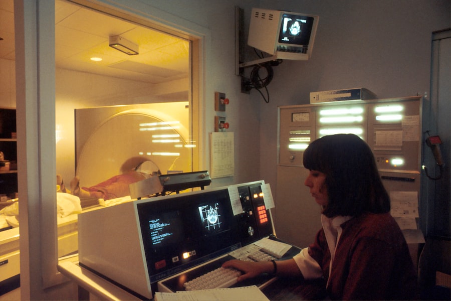
**Imaging Studies**: Magnetic resonance imaging (MRI) is the gold standard for visualizing brain tumors. It provides detailed images of brain structures and helps determine tumor size, location, and potential involvement of surrounding tissues. 2.
**Neurological Assessment**: A comprehensive neurological examination assesses motor function, sensory perception, coordination, reflexes, and cognitive abilities to identify any deficits that may indicate tumor presence. 3. **Biopsy**: In many cases, a biopsy is necessary to confirm the diagnosis of glioma.
This procedure involves obtaining a tissue sample from the tumor for histopathological analysis. Early diagnosis is critical for effective management and treatment planning for patients with glioma brain tumors.
Imaging Tests for Glioma Brain Tumors
Common Imaging Modalities
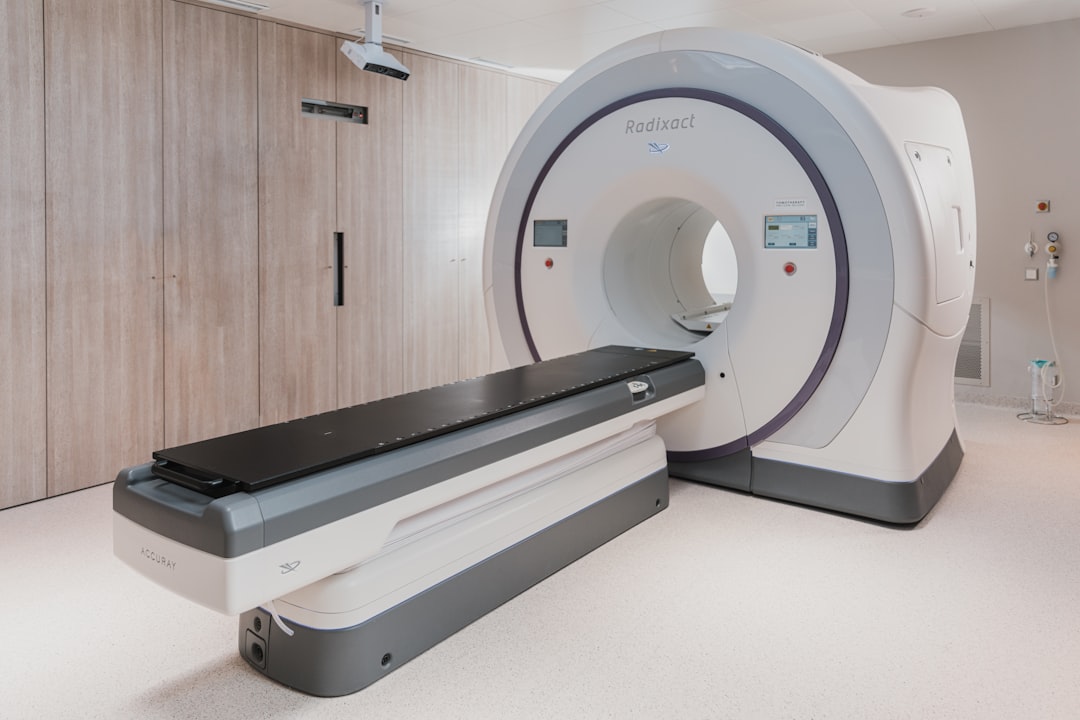
The most commonly used imaging modalities include Magnetic Resonance Imaging (MRI), Computed Tomography (CT) Scan, Functional MRI (fMRI), and Positron Emission Tomography (PET) Scan.
It is particularly useful for assessing tumor size, location, and any associated edema or mass effect on surrounding tissues. CT scans, while less sensitive than MRI for detecting brain tumors, can be useful in emergency settings or when MRI is contraindicated. They can quickly identify significant mass effects or hemorrhage. fMRI assesses brain activity by measuring changes in blood flow associated with neuronal activity, helping to identify critical functional areas of the brain before surgery. PET scans utilize radioactive tracers to visualize metabolic activity within the tumor, helping to differentiate between tumor recurrence and radiation necrosis in post-treatment evaluations.
Importance in Diagnosis and Treatment
These imaging techniques provide essential information that aids in accurate diagnosis and treatment planning for glioma patients.
Biopsy and Pathology for Glioma Brain Tumors
A biopsy is often necessary to confirm the diagnosis of a glioma brain tumor definitively. There are several methods for obtaining tissue samples: 1. **Stereotactic Biopsy**: This minimally invasive procedure uses advanced imaging guidance to precisely target the tumor location for tissue sampling.
It is particularly useful for deep-seated tumors that are difficult to access surgically. 2. **Open Biopsy**: In some cases, an open biopsy may be performed during surgical resection of the tumor.
This approach allows for larger tissue samples to be obtained for comprehensive histopathological analysis.
Accurate pathology results are crucial for determining prognosis and guiding treatment decisions for patients diagnosed with gliomas.
Treatment Options for Glioma Brain Tumors
The management of glioma brain tumors typically involves a multidisciplinary approach tailored to each patient’s unique circumstances. Treatment options may include: 1. **Surgery**: Surgical resection aims to remove as much tumor tissue as possible while preserving surrounding healthy brain tissue.
Complete resection is often associated with improved outcomes; however, it may not always be feasible due to tumor location or infiltration into critical brain areas. 2. **Radiation Therapy**: Radiation therapy is commonly employed post-surgery to target residual tumor cells and reduce the risk of recurrence.
Techniques such as external beam radiation therapy (EBRT) or stereotactic radiosurgery (SRS) may be utilized based on tumor characteristics. 3. **Chemotherapy**: Chemotherapy agents such as temozolomide (TMZ) are frequently used in conjunction with radiation therapy for high-grade gliomas like glioblastoma multiforme.
Chemotherapy aims to target rapidly dividing tumor cells while minimizing damage to normal cells. 4. **Targeted Therapy**: Emerging therapies targeting specific molecular pathways involved in glioma growth are being investigated in clinical trials.
These therapies aim to inhibit tumor progression while sparing healthy tissue. The choice of treatment depends on various factors including tumor type, grade, location, patient age, and overall health status.
Surgery for Glioma Brain Tumors
Surgical intervention remains a cornerstone in the management of glioma brain tumors when feasible. The primary goals of surgery include: 1. **Maximal Safe Resection**: The objective is to remove as much tumor tissue as possible while preserving critical neurological function.
Surgeons utilize advanced techniques such as intraoperative MRI or neuronavigation systems to enhance precision during resection. 2. **Debulking Surgery**: In cases where complete resection is not achievable due to tumor location or infiltration into vital structures, debulking surgery may be performed to reduce tumor burden and alleviate symptoms associated with increased intracranial pressure.
3. **Palliative Surgery**: In certain situations where curative intent is not possible, palliative surgical procedures may be performed to relieve symptoms or improve quality of life. Postoperative care is essential for monitoring neurological function and managing potential complications such as infection or seizures.
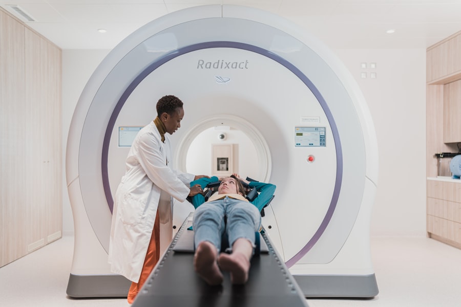
Radiation Therapy for Glioma Brain Tumors
Radiation therapy plays a critical role in the management of glioma brain tumors, particularly following surgical resection: 1. **Adjuvant Radiation Therapy**: After surgery, adjuvant radiation therapy is often recommended to target residual tumor cells that may not have been removed during surgery. This approach aims to reduce the risk of recurrence and improve overall survival rates.
2. **Techniques Used**: External beam radiation therapy (EBRT) delivers targeted radiation beams to the tumor site while minimizing exposure to surrounding healthy tissue. Stereotactic radiosurgery (SRS) offers a non-invasive alternative that delivers high doses of radiation precisely to the tumor while sparing adjacent structures.
3. **Side Effects Management**: Common side effects of radiation therapy include fatigue, skin irritation at the treatment site, and potential cognitive changes over time. Supportive care measures are essential to manage these side effects effectively.
Radiation therapy remains a vital component in the comprehensive management of glioma patients.
Chemotherapy and Targeted Therapy for Glioma Brain Tumors
Chemotherapy has become an integral part of treatment protocols for high-grade gliomas: 1. **Temozolomide (TMZ)**: This oral chemotherapy agent is commonly used in conjunction with radiation therapy for patients diagnosed with glioblastoma multiforme (GBM). TMZ works by damaging DNA in rapidly dividing cancer cells, ultimately leading to cell death.
2. **Adverse Effects**: While effective, chemotherapy can lead to side effects such as nausea, vomiting, fatigue, hair loss, and myelosuppression (decreased bone marrow function). Regular monitoring of blood counts is essential during treatment.
3. **Targeted Therapies**: Research into targeted therapies aims to exploit specific molecular alterations present in gliomas. For instance, inhibitors targeting mutations in the IDH1 gene or therapies aimed at blocking angiogenesis (the formation of new blood vessels) are under investigation in clinical trials.
The landscape of chemotherapy and targeted therapy continues to evolve as new agents are developed and tested in clinical settings. In summary, glioma brain tumors represent a complex group of neoplasms that require careful diagnosis and management through a multidisciplinary approach involving surgery, radiation therapy, chemotherapy, and emerging targeted therapies tailored to individual patient needs. Understanding the various types of gliomas, their symptoms, risk factors, diagnostic methods, treatment options, and ongoing research efforts is crucial for improving patient outcomes in this challenging field of neuro-oncology.
There have been significant advancements in the treatment of glioma brain tumors, with researchers constantly striving to improve outcomes for patients. One related article discusses the importance of early detection and personalized treatment options for glioma patients. To learn more about this topic, check out this article on the Boogger website.
FAQs
What is a glioma brain tumor?
A glioma is a type of tumor that starts in the glial cells of the brain or the spine. Glial cells are supportive cells that help keep the neurons in the brain functioning properly.
What are the symptoms of a glioma brain tumor?
Symptoms of a glioma brain tumor can vary depending on the location and size of the tumor, but common symptoms may include headaches, seizures, nausea, vomiting, changes in vision or hearing, and difficulty with balance or walking.
How is a glioma brain tumor diagnosed?
Diagnosis of a glioma brain tumor typically involves a combination of imaging tests such as MRI or CT scans, as well as a biopsy to examine the tumor tissue under a microscope.
What are the treatment options for glioma brain tumors?
Treatment options for glioma brain tumors may include surgery to remove the tumor, radiation therapy, and chemotherapy. The specific treatment plan will depend on the type and grade of the tumor, as well as the patient’s overall health.
What is the prognosis for glioma brain tumors?
The prognosis for glioma brain tumors can vary widely depending on factors such as the type and grade of the tumor, the age and overall health of the patient, and how well the tumor responds to treatment. It is important for patients to work closely with their healthcare team to develop a personalized treatment plan.