Now Reading: Nodular Basal Cell Carcinoma: What You Need to Know
-
01
Nodular Basal Cell Carcinoma: What You Need to Know
Nodular Basal Cell Carcinoma: What You Need to Know
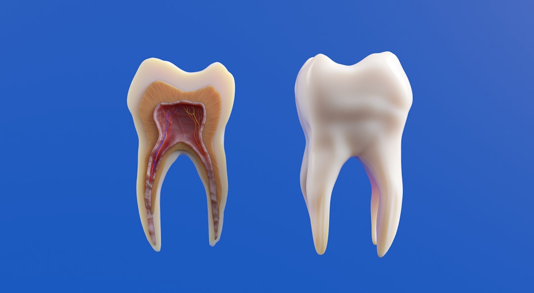
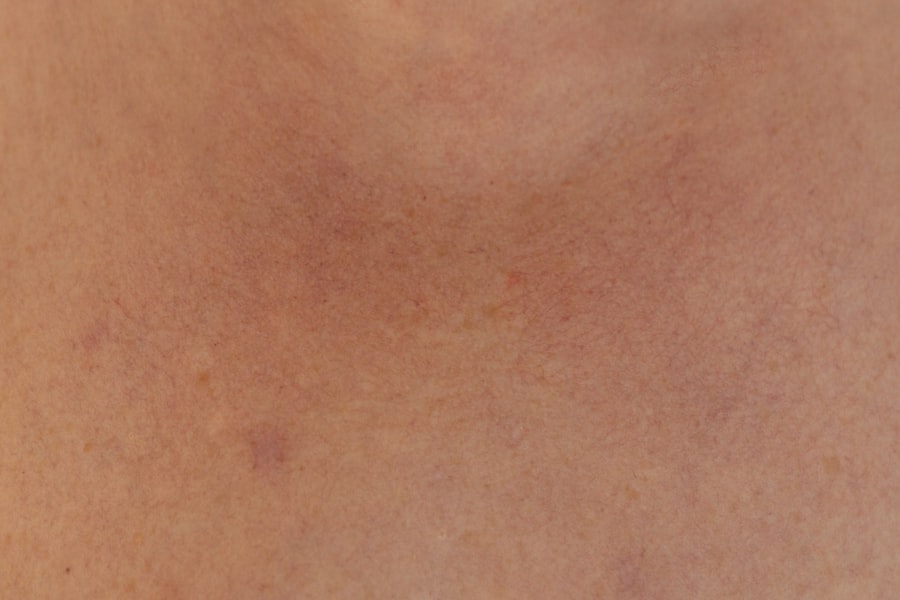
Nodular Basal Cell Carcinoma (NBCC) is the most prevalent form of skin cancer, originating from the basal cells located in the epidermis, the outermost layer of the skin. This malignancy is characterized by its slow growth and typically manifests as a pearly or waxy bump on sun-exposed areas, particularly the face, ears, neck, and scalp. While it is generally considered less aggressive than other skin cancers, such as melanoma, it can lead to significant local tissue destruction if left untreated.
The tumor’s indolent nature often results in a favorable prognosis; however, early detection and intervention are crucial to prevent complications. The histological features of NBCC include nests of basaloid cells with peripheral palisading and a stroma that may exhibit varying degrees of desmoplasia. The tumor’s growth pattern can be infiltrative, nodular, or superficial, with the nodular variant being the most common.
Understanding the biological behavior of this carcinoma is essential for effective management and treatment strategies.
Key Takeaways
- Nodular basal cell carcinoma is a common type of skin cancer that appears as a flesh-colored or pink bump on the skin.
- Causes and risk factors for nodular basal cell carcinoma include excessive sun exposure, tanning bed use, and a family history of skin cancer.
- Signs and symptoms of nodular basal cell carcinoma may include a shiny or waxy bump, a bleeding or oozing sore, or a scar-like area.
- Diagnosis and tests for nodular basal cell carcinoma may involve a skin biopsy or a dermatoscopy to examine the skin lesion more closely.
- Treatment options for nodular basal cell carcinoma include surgical procedures such as excision, Mohs surgery, and non-surgical options like cryotherapy and topical medications.
Causes and Risk Factors
The primary etiological factor for nodular basal cell carcinoma is prolonged exposure to ultraviolet (UV) radiation from sunlight. This exposure leads to DNA damage in skin cells, which can initiate carcinogenesis. Individuals with fair skin, light-colored eyes, and red or blonde hair are particularly susceptible due to lower levels of melanin, which provides some protection against UV radiation.
Other significant risk factors include a history of sunburns, especially during childhood, and living in sunny climates. Additionally, individuals with compromised immune systems, such as organ transplant recipients or those with certain genetic conditions like Gorlin syndrome, are at an increased risk for developing NBCChronic exposure to certain chemicals, such as arsenic or coal tar, has also been implicated in the pathogenesis of this skin cancer.
Signs and Symptoms
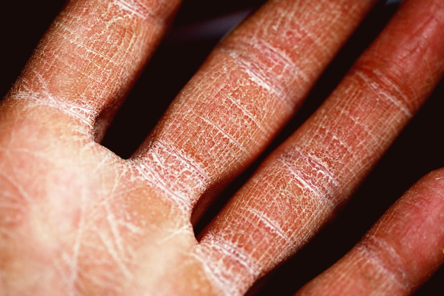
Nodular basal cell carcinoma typically presents as a small, shiny bump that may be translucent or pearly in appearance. These lesions can vary in color from skin-toned to pink or even brownish. As the tumor progresses, it may develop a central ulceration or crusting, leading to a non-healing sore that can bleed or ooze.
Patients may also notice changes in the texture of the skin surrounding the lesion, which can become scaly or rough. In some cases, NBCC may be asymptomatic; however, patients often report itching or tenderness in the affected area. It is essential to differentiate these lesions from other dermatological conditions such as seborrheic keratosis or actinic keratosis to ensure appropriate management.
Regular self-examinations and awareness of any changes in the skin are vital for early detection.
Diagnosis and Tests
| Diagnosis and Tests | Accuracy | Cost |
|---|---|---|
| Blood Test | High | Low |
| CT Scan | High | High |
| MRI | High | High |
| Ultrasound | Medium | Low |
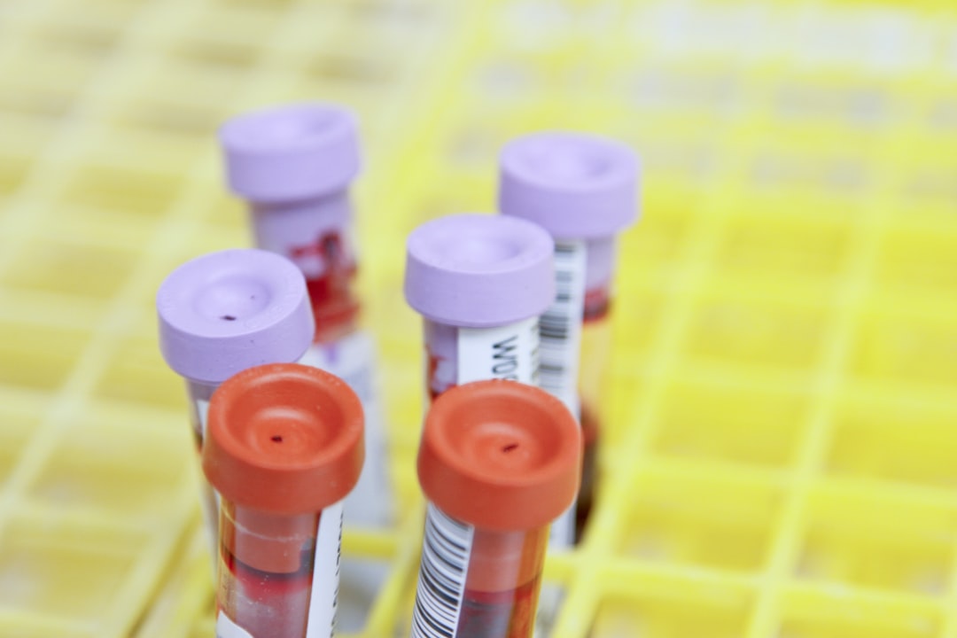
The diagnosis of nodular basal cell carcinoma primarily involves a thorough clinical examination by a dermatologist. The physician will assess the characteristics of the lesion and may perform a dermatoscopic examination to evaluate the lesion’s structure more closely. If there is suspicion of malignancy, a biopsy is typically performed to confirm the diagnosis.
There are several types of biopsies that may be utilized, including shave biopsy, punch biopsy, or excisional biopsy.
Histopathological examination of the biopsy specimen will reveal characteristic features of NBCC, including nests of basaloid cells and peripheral palisading.
In some cases, imaging studies such as ultrasound or MRI may be employed to assess for deeper invasion or metastasis, although this is rare for NBCC.
Treatment Options
Treatment for nodular basal cell carcinoma is primarily determined by factors such as tumor size, location, and patient health status. The main goal is to achieve complete excision of the tumor while preserving surrounding healthy tissue. Surgical excision remains the gold standard for treatment and involves removing the tumor along with a margin of healthy skin to minimize recurrence risk.
In addition to surgical options, various non-surgical treatments are available for patients who may not be candidates for surgery or prefer alternative approaches. These include topical chemotherapy agents like 5-fluorouracil (5-FU) and imiquimod, which stimulate an immune response against cancer cells. Photodynamic therapy (PDT) is another option that utilizes light-activated drugs to destroy cancerous cells.
Surgical Procedures
Surgical excision is often performed under local anesthesia and involves removing the tumor along with a margin of healthy tissue. Mohs micrographic surgery is a specialized technique that allows for real-time examination of excised tissue margins during surgery. This method ensures complete removal of cancerous cells while preserving as much healthy tissue as possible.
Other surgical options include curettage and electrodessication, where the tumor is scraped away and then cauterized to destroy any remaining cancerous cells. Cryotherapy may also be employed in select cases, where liquid nitrogen is used to freeze and destroy abnormal cells. Each surgical approach has its indications based on tumor characteristics and patient preferences.
Non-Surgical Treatment Options
For patients who are not suitable candidates for surgery due to health concerns or those who prefer non-invasive methods, several non-surgical treatment options exist. Topical chemotherapy agents like 5-fluorouracil (5-FU) are applied directly to the lesion over several weeks to induce localized destruction of cancerous cells. Imiquimod cream works by stimulating the immune system to attack cancer cells and is particularly effective for superficial forms of basal cell carcinoma.
Photodynamic therapy (PDT) involves applying a photosensitizing agent to the lesion followed by exposure to a specific wavelength of light that activates the drug, leading to selective destruction of cancerous cells. This method is less invasive and can be performed in an outpatient setting.
Potential Complications
While nodular basal cell carcinoma has a high cure rate when treated appropriately, potential complications can arise if left untreated or inadequately managed. Local invasion can lead to significant tissue destruction, particularly in cosmetically sensitive areas such as the face or ears. In rare cases, aggressive variants of basal cell carcinoma can metastasize to distant sites, although this occurrence is exceedingly uncommon.
Post-treatment complications may include scarring or changes in pigmentation at the site of excision or treatment. Patients should be counseled on proper wound care following surgical procedures to minimize these risks and promote optimal healing.
Prevention and Early Detection
Preventive measures play a crucial role in reducing the incidence of nodular basal cell carcinoma. Limiting sun exposure during peak hours (10 AM to 4 PM), wearing protective clothing, and using broad-spectrum sunscreen with an SPF of 30 or higher are essential strategies for safeguarding against UV damage. Regular skin examinations by a dermatologist can facilitate early detection of suspicious lesions.
Self-examinations should be conducted monthly, focusing on any new growths or changes in existing moles or spots on the skin. Education on recognizing early signs of skin cancer can empower individuals to seek timely medical attention.
Prognosis and Outlook
The prognosis for patients diagnosed with nodular basal cell carcinoma is generally favorable due to its slow-growing nature and low metastatic potential. When detected early and treated appropriately, the five-year survival rate approaches 100%. However, recurrence can occur in some cases, particularly if margins are not adequately cleared during surgical excision.
Long-term follow-up is recommended for patients with a history of NBCC due to their increased risk for developing new skin cancers over time. Regular dermatological evaluations can help monitor for any new lesions or recurrences.
Support and Resources
Patients diagnosed with nodular basal cell carcinoma may benefit from various support resources available through cancer organizations and local support groups. The American Cancer Society provides comprehensive information on skin cancer prevention, treatment options, and emotional support resources for patients and their families. Additionally, online platforms offer forums where individuals can share experiences and coping strategies related to their diagnosis and treatment journey.
Engaging with healthcare professionals who specialize in dermatology and oncology can also provide valuable insights into managing this condition effectively. In summary, nodular basal cell carcinoma represents a common yet manageable form of skin cancer characterized by its slow growth and favorable prognosis when treated early. Understanding its causes, risk factors, signs, symptoms, diagnostic methods, treatment options—including both surgical and non-surgical approaches—can empower individuals to take proactive steps toward prevention and early detection.
Regular follow-ups and support resources play an essential role in ensuring optimal outcomes for patients navigating this diagnosis.
Nodular basal cell carcinoma is a common type of skin cancer that often appears as a shiny bump on the skin. According to a recent article on boogger.com, early detection and treatment of nodular basal cell carcinoma is crucial in preventing further complications. It is important to regularly check your skin for any changes and consult a dermatologist if you notice any suspicious growths.
FAQs
What is nodular basal cell carcinoma?
Nodular basal cell carcinoma is a type of skin cancer that arises from the basal cells in the skin’s outermost layer. It typically appears as a firm, red or flesh-colored nodule on the skin.
What are the symptoms of nodular basal cell carcinoma?
Symptoms of nodular basal cell carcinoma may include a shiny or pearly bump, a pink growth with slightly raised borders, a scar-like area that is white, yellow, or waxy, or a bleeding or oozing sore that doesn’t heal.
How is nodular basal cell carcinoma diagnosed?
Nodular basal cell carcinoma is diagnosed through a skin biopsy, where a small sample of the affected skin is removed and examined under a microscope by a dermatologist or pathologist.
What are the treatment options for nodular basal cell carcinoma?
Treatment options for nodular basal cell carcinoma may include surgical excision, Mohs surgery, cryotherapy, radiation therapy, and topical medications such as imiquimod or 5-fluorouracil.
What are the risk factors for nodular basal cell carcinoma?
Risk factors for nodular basal cell carcinoma include prolonged sun exposure, a history of sunburns, a family history of skin cancer, fair skin, and a weakened immune system.
Can nodular basal cell carcinoma be prevented?
Nodular basal cell carcinoma can be prevented by practicing sun safety measures such as wearing sunscreen, protective clothing, and seeking shade, as well as avoiding tanning beds and sunlamps. Regular skin checks and early detection are also important for prevention.