Now Reading: Pseudomyxoma Peritonei: Understanding the Rare Abdominal Condition
-
01
Pseudomyxoma Peritonei: Understanding the Rare Abdominal Condition
Pseudomyxoma Peritonei: Understanding the Rare Abdominal Condition

Pseudomyxoma peritonei (PMP) is a rare and complex condition characterized by the accumulation of mucinous material within the peritoneal cavity, often resulting from a neoplastic process. This condition typically arises from appendiceal tumors, particularly low-grade appendiceal mucinous neoplasms (LAMNs), which can lead to the production of excessive mucin. The term “pseudomyxoma” refers to the gelatinous appearance of the mucinous fluid that fills the abdominal cavity, mimicking a true mucinous tumor.
PMP is not classified as a primary cancer but rather as a complication of a neoplastic process, often leading to significant morbidity due to its progressive nature. The pathophysiology of PMP involves the dissemination of mucin-producing cells throughout the peritoneal cavity, leading to the formation of mucinous ascites. This accumulation can cause abdominal distension, discomfort, and various gastrointestinal symptoms.
The condition is often insidious in onset, with patients frequently experiencing vague symptoms that can delay diagnosis. Understanding the nature of PMP is crucial for effective management and treatment, as it requires a multidisciplinary approach involving surgical intervention and potentially adjuvant therapies.
Key Takeaways
- Pseudomyxoma Peritonei is a rare cancer that usually starts in the appendix and can spread to the lining of the abdominal cavity.
- Causes and risk factors of Pseudomyxoma Peritonei include a ruptured appendix, genetic predisposition, and certain medical conditions.
- Signs and symptoms of Pseudomyxoma Peritonei may include abdominal pain, bloating, changes in bowel habits, and unexplained weight loss.
- Diagnosing Pseudomyxoma Peritonei involves imaging tests, blood tests, and a biopsy to confirm the presence of mucinous tumors in the abdomen.
- Treatment options for Pseudomyxoma Peritonei may include surgery, chemotherapy, and heated intraperitoneal chemotherapy (HIPEC) to remove and destroy cancerous cells.
Causes and Risk Factors of Pseudomyxoma Peritonei
The primary cause of pseudomyxoma peritonei is the rupture of an appendiceal tumor, particularly low-grade appendiceal mucinous neoplasms. These tumors can produce large quantities of mucin, which, when released into the peritoneal cavity, leads to the characteristic accumulation seen in PMP.
Genetic predispositions, such as familial adenomatous polyposis (FAP) or Lynch syndrome, may also increase the risk of developing appendiceal tumors that can lead to PMP. Several risk factors have been identified that may contribute to the development of PMP. These include age, with most cases occurring in individuals between 40 and 70 years old, and gender, as males are more frequently affected than females.
Additionally, a history of appendicitis or previous abdominal surgeries may predispose individuals to PMP due to potential disruptions in normal anatomical barriers. Understanding these risk factors is essential for early detection and intervention.
Signs and Symptoms of Pseudomyxoma Peritonei
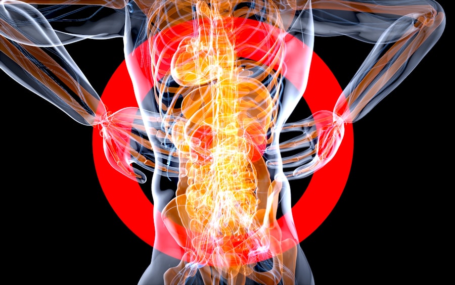
Patients with pseudomyxoma peritonei often present with a range of nonspecific symptoms that can complicate diagnosis. Common manifestations include abdominal distension, pain, and discomfort due to the accumulation of mucinous fluid. Patients may also experience changes in bowel habits, such as constipation or diarrhea, as well as early satiety due to pressure on the gastrointestinal tract.
In some cases, patients may report weight loss or fatigue, which can be attributed to the chronic nature of the condition. As PMP progresses, more severe symptoms may develop, including bowel obstruction or perforation due to the mass effect of the mucinous material. Ascites may become more pronounced, leading to significant abdominal swelling and discomfort.
In advanced cases, patients may experience systemic symptoms such as fever or malaise, indicating potential complications or secondary infections.
Diagnosing Pseudomyxoma Peritonei
| Diagnosis Method | Accuracy |
|---|---|
| Imaging tests (CT, MRI) | 80% |
| Biopsy | 70% |
| Tumor markers (CEA, CA 19-9) | 60% |
The diagnosis of pseudomyxoma peritonei typically involves a combination of imaging studies and histopathological evaluation. Initial imaging modalities include ultrasound and computed tomography (CT) scans, which can reveal characteristic findings such as loculated ascites and thickened peritoneal surfaces. Magnetic resonance imaging (MRI) may also be utilized for further characterization of the disease.
Once imaging suggests the presence of PMP, a definitive diagnosis often requires histological confirmation through biopsy. This may involve laparoscopic or open surgical techniques to obtain tissue samples from affected areas within the peritoneal cavity. Pathological examination reveals mucinous material and atypical cells consistent with low-grade appendiceal mucinous neoplasms.
Accurate diagnosis is essential for determining appropriate treatment strategies and prognostic implications.
Treatment Options for Pseudomyxoma Peritonei
The management of pseudomyxoma peritonei primarily revolves around surgical intervention, as this condition is not amenable to conventional chemotherapy or radiation therapy alone. The cornerstone of treatment is cytoreductive surgery (CRS), which aims to remove all visible tumor deposits from the peritoneal cavity. This procedure is often combined with hyperthermic intraperitoneal chemotherapy (HIPEC), where heated chemotherapy agents are delivered directly into the abdominal cavity post-surgery to target residual microscopic disease.
In cases where complete cytoreduction is not feasible due to extensive disease burden or patient comorbidities, palliative measures may be considered. These can include repeated paracentesis to relieve symptomatic ascites or placement of intraperitoneal catheters for continuous drainage. Additionally, clinical trials exploring novel therapeutic agents and immunotherapy are ongoing, providing hope for improved outcomes in patients with advanced PMP.
Surgical Procedures for Pseudomyxoma Peritonei

Cytoreductive surgery for pseudomyxoma peritonei is a complex procedure that requires meticulous planning and execution by a skilled surgical team. The goal is to achieve optimal cytoreduction by excising all visible tumor masses while preserving vital organs and structures within the abdomen. This may involve resection of affected organs such as the appendix, portions of the colon, or ovaries if involved.
Following cytoreduction, hyperthermic intraperitoneal chemotherapy (HIPEC) is administered to enhance local control of disease. During HIPEC, chemotherapy agents such as cisplatin or mitomycin C are heated to approximately 42 degrees Celsius and circulated within the abdominal cavity for a specified duration. This approach aims to maximize drug penetration into tumor tissues while minimizing systemic toxicity.
The combination of CRS and HIPEC has been associated with improved survival rates in select patient populations.
Follow-Up Care and Monitoring for Pseudomyxoma Peritonei
Postoperative follow-up care is critical for patients who have undergone treatment for pseudomyxoma peritonei. Regular monitoring through clinical evaluations and imaging studies is essential to detect any recurrence or progression of disease early on. Follow-up schedules typically involve visits every three to six months during the first two years post-treatment, transitioning to annual assessments thereafter if no evidence of disease is present.
Patients should also be educated about potential late effects of treatment, including bowel dysfunction or changes in nutritional status due to surgical interventions. Nutritional support may be necessary for those experiencing difficulties with food intake or absorption. Psychological support services should also be considered to address any emotional challenges faced by patients during their recovery journey.
Living with Pseudomyxoma Peritonei: Coping and Support
Living with pseudomyxoma peritonei can be challenging both physically and emotionally for patients and their families. Coping strategies may include engaging in support groups where individuals can share experiences and gain insights from others facing similar challenges. These groups provide a platform for emotional expression and foster a sense of community among patients navigating their journeys.
Additionally, mental health professionals specializing in oncology can offer valuable support through counseling or therapy sessions tailored to address anxiety, depression, or fear related to diagnosis and treatment outcomes. Encouraging open communication with healthcare providers about concerns or symptoms can empower patients in their care journey and enhance their overall quality of life.
Research and Advances in Pseudomyxoma Peritonei
Ongoing research into pseudomyxoma peritonei aims to improve understanding of its pathogenesis and develop more effective treatment modalities. Investigations into genetic markers associated with appendiceal tumors are underway, potentially leading to targeted therapies that could enhance patient outcomes. Furthermore, clinical trials exploring novel chemotherapeutic agents and immunotherapy approaches are being conducted to assess their efficacy in managing PMP.
Advancements in surgical techniques and technologies also hold promise for improving surgical outcomes in patients with PMP. Minimally invasive approaches are being evaluated for their potential benefits in reducing postoperative complications and recovery times while maintaining oncological efficacy.
Prognosis and Outlook for Pseudomyxoma Peritonei
The prognosis for patients with pseudomyxoma peritonei varies significantly based on several factors, including tumor grade, extent of disease at diagnosis, and completeness of cytoreduction achieved during surgery. Generally, patients who undergo successful cytoreductive surgery followed by HIPEC have better survival rates compared to those who do not receive aggressive treatment. Long-term survival rates can range from 30% to over 70% at five years post-treatment for those with low-grade disease who achieve complete cytoreduction.
However, patients with high-grade tumors or incomplete cytoreduction may have a more guarded prognosis. Continuous follow-up care is essential for monitoring disease recurrence and managing any complications that may arise.
Resources and Support for Pseudomyxoma Peritonei Patients and Caregivers
Numerous resources are available for patients diagnosed with pseudomyxoma peritonei and their caregivers. Organizations such as the Pseudomyxoma Peritonei Research Foundation provide educational materials, support networks, and information on clinical trials aimed at advancing research in this area. Additionally, online forums and social media groups offer platforms for individuals to connect with others facing similar challenges.
Healthcare providers play a crucial role in guiding patients toward appropriate resources for emotional support, nutritional counseling, and rehabilitation services post-treatment. Engaging with multidisciplinary teams ensures comprehensive care that addresses both physical and psychosocial aspects of living with pseudomyxoma peritonei. In summary, pseudomyxoma peritonei is a rare condition resulting from mucinous tumors primarily originating from the appendix.
Early diagnosis through imaging studies and histopathological evaluation is critical for effective management. Treatment typically involves cytoreductive surgery combined with HIPEC; however, ongoing research continues to explore novel therapeutic options. Patients must engage in regular follow-up care while utilizing available resources for emotional support throughout their journey with this complex condition.
Pseudomyxoma peritonei is a rare type of cancer that usually starts in the appendix and spreads to the peritoneum. For more information on navigating the best rates for car insurance in 2025, check out this article. It is important to stay informed about different topics, including federal holidays in August, which you can learn more about in this guide.
FAQs
What is pseudomyxoma peritonei?
Pseudomyxoma peritonei is a rare type of cancer that usually starts in the appendix and can spread to the peritoneum, which is the lining of the abdominal cavity.
What are the symptoms of pseudomyxoma peritonei?
Symptoms of pseudomyxoma peritonei may include abdominal pain or discomfort, bloating, changes in bowel habits, and unexplained weight loss.
How is pseudomyxoma peritonei diagnosed?
Pseudomyxoma peritonei is typically diagnosed through imaging tests such as CT scans, MRI scans, and ultrasound, as well as through a biopsy to examine the tissue for cancer cells.
What are the treatment options for pseudomyxoma peritonei?
Treatment for pseudomyxoma peritonei often involves a combination of surgery to remove the tumor and chemotherapy to kill any remaining cancer cells. In some cases, heated intraperitoneal chemotherapy (HIPEC) may also be used.
What is the prognosis for pseudomyxoma peritonei?
The prognosis for pseudomyxoma peritonei can vary depending on the stage of the cancer and the individual’s overall health. However, with appropriate treatment, many people with pseudomyxoma peritonei can live for many years after diagnosis.