Now Reading: Understanding Lung Cancer Stages
-
01
Understanding Lung Cancer Stages
Understanding Lung Cancer Stages
Lung cancer is classified into distinct stages that reflect the extent of the disease and its progression. The most widely accepted staging system is the TNM classification, which evaluates three primary components: Tumor size (T), lymph Node involvement (N), and Metastasis (M). Lung cancer is generally categorized into four main stages, ranging from Stage 0 to Stage
Key Takeaways
- Lung cancer stages range from 0 to IV, with stage 0 being the earliest and stage IV being the most advanced.
- Understanding lung cancer stages is crucial for determining the appropriate treatment plan and predicting the patient’s prognosis.
- Lung cancer stages are determined based on the size of the tumor, whether it has spread to nearby lymph nodes, and if it has metastasized to other parts of the body.
- Non-small cell lung cancer and small cell lung cancer have different staging systems, with non-small cell lung cancer using the TNM system and small cell lung cancer using limited or extensive stage classification.
- Lung cancer stages greatly impact treatment options, with earlier stages often being treated with surgery and later stages requiring a combination of chemotherapy, radiation, and targeted therapy.
- The prognosis for each stage of lung cancer varies, with higher survival rates for earlier stages and lower survival rates for advanced stages.
- The spread of lung cancer to nearby lymph nodes or distant organs can increase the stage of the cancer, indicating a poorer prognosis and more aggressive treatment.
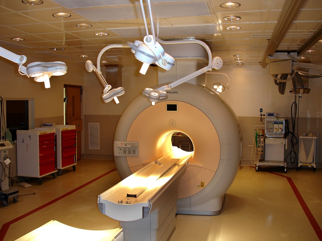
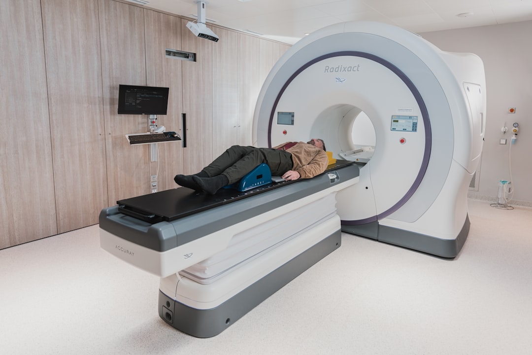
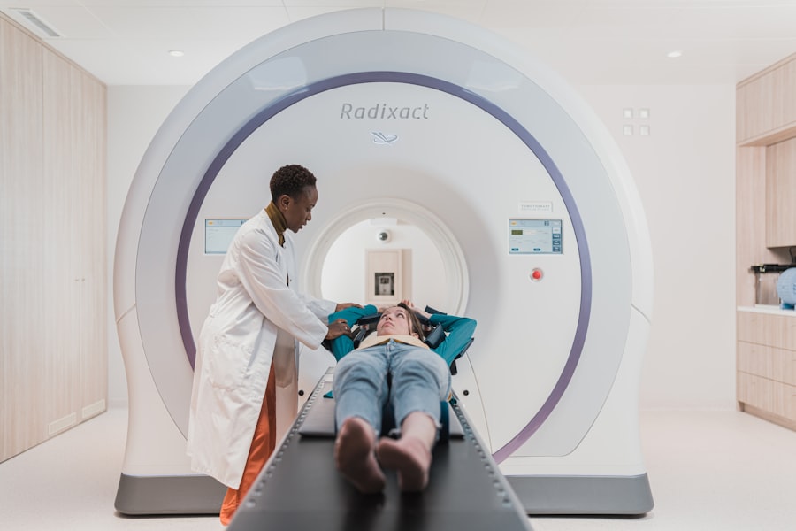
- Imaging tests such as CT scans, PET scans, and MRI are essential in determining the stage of lung cancer by providing detailed information about the size and location of the tumor, as well as any spread to other parts of the body.
- The TNM staging system for lung cancer evaluates the size and extent of the primary tumor (T), the involvement of nearby lymph nodes (N), and the presence of distant metastasis (M).
- The stage of lung cancer significantly affects survival rates, with earlier stages having higher survival rates and advanced stages having lower survival rates.
- Being diagnosed with a specific stage of lung cancer can have a profound emotional impact on patients and their families, influencing their outlook on the disease and their treatment decisions.
– **Stage 0**: Also known as carcinoma in situ, this stage indicates that abnormal cells are present in the lining of the lung but have not invaded deeper tissues or spread to lymph nodes.
– **Stage I**: At this stage, the tumor is localized within the lung and has not spread to nearby lymph nodes. It is further divided into IA and IB based on tumor size and specific characteristics.
– **Stage II**: This stage signifies that the cancer has grown larger and may have spread to nearby lymph nodes. It is also subdivided into IIA and IIB, depending on the extent of lymph node involvement.
– **Stage III**: This stage indicates more extensive disease, with cancer having spread to lymph nodes in the mediastinum or chest wall.
It is categorized into IIIA and IIIB based on the extent of spread.
– **Stage IV**: The most advanced stage, where cancer has metastasized to distant organs such as the liver, brain, or bones. This stage is further classified into IVA and IVB based on the number and location of metastases. Understanding these stages is crucial for determining treatment options and predicting outcomes.
The importance of understanding lung cancer stages
Comprehending the stages of lung cancer is vital for both patients and healthcare providers. Accurate staging allows for tailored treatment plans that can significantly influence patient outcomes. For instance, early-stage lung cancer (Stages 0 and I) may be amenable to surgical resection, while advanced stages (III and IV) often require a combination of chemotherapy, radiation therapy, and targeted therapies.
Moreover, staging provides insight into prognosis. Patients diagnosed at earlier stages generally have a better survival rate compared to those diagnosed at later stages. Understanding the stage also helps in setting realistic expectations regarding treatment efficacy and potential side effects.
Additionally, knowledge of lung cancer stages can empower patients to make informed decisions about their care. It fosters open communication between patients and healthcare providers, enabling discussions about clinical trials, palliative care options, and lifestyle modifications that may enhance quality of life.
How are lung cancer stages determined?
| Stage | Description |
|---|---|
| Stage 0 | Cancer is only in the top layer of cells lining the air passages and has not spread to deeper lung tissue. |
| Stage I | Cancer is in the lung but has not spread to the lymph nodes. |
| Stage II | Cancer is in the lung and nearby lymph nodes. |
| Stage III | Cancer is in the lung and lymph nodes in the middle of the chest. |
| Stage IV | Cancer has spread to both lungs, into the area around the lungs, or to distant organs. |
The determination of lung cancer stages involves a comprehensive evaluation that includes medical history, physical examinations, imaging studies, and sometimes invasive procedures. Initially, a thorough medical history is taken to assess risk factors such as smoking history, exposure to carcinogens, and family history of lung cancer. Imaging tests play a crucial role in staging.
Chest X-rays, CT scans, PET scans, and MRI scans are commonly employed to visualize the lungs and surrounding structures. These imaging modalities help identify tumor size, location, and any potential metastases. In some cases, a biopsy may be necessary to confirm the diagnosis and assess tumor characteristics.
This procedure involves obtaining a sample of lung tissue for histopathological examination. The results from imaging studies and biopsies are then integrated into the TNM classification system to assign an accurate stage.
The difference between non-small cell lung cancer and small cell lung cancer stages
Lung cancer is primarily categorized into two main types: non-small cell lung cancer (NSCLC) and small cell lung cancer (SCLC). Each type has distinct staging criteria due to differences in biology and behavior. Non-small cell lung cancer accounts for approximately 85% of all lung cancer cases and includes subtypes such as adenocarcinoma, squamous cell carcinoma, and large cell carcinoma.
The staging for NSCLC follows the TNM system closely, allowing for detailed assessment based on tumor size, lymph node involvement, and metastasis. Conversely, small cell lung cancer is more aggressive and tends to spread rapidly. SCLC is typically classified into two stages: limited stage (where cancer is confined to one lung and nearby lymph nodes) and extensive stage (where cancer has spread beyond the original site).
This simplified staging reflects the urgent need for aggressive treatment strategies due to the rapid progression associated with SCLC.
The impact of lung cancer stages on treatment options

The stage of lung cancer significantly influences treatment decisions. For early-stage NSCLC (Stages 0 and I), surgical resection is often the primary treatment modality. Lobectomy or pneumonectomy may be performed to remove the tumor along with surrounding healthy tissue.
Adjuvant chemotherapy may be recommended post-surgery to reduce recurrence risk. In Stage II NSCLC, treatment may involve a combination of surgery followed by chemotherapy or radiation therapy to target any residual disease. For Stage III NSCLC, a multimodal approach is typically employed, combining chemotherapy, radiation therapy, and possibly surgery depending on individual patient factors.
For advanced Stage IV lung cancer, treatment focuses on palliative care aimed at managing symptoms and improving quality of life. Targeted therapies such as tyrosine kinase inhibitors or immune checkpoint inhibitors may be utilized based on specific genetic mutations present in the tumor. In contrast, small cell lung cancer treatment primarily involves chemotherapy and radiation therapy due to its aggressive nature.
Limited-stage SCLC may be treated with concurrent chemoradiation, while extensive-stage SCLC often requires systemic chemotherapy as the mainstay of treatment.
The prognosis for each stage of lung cancer
Prognosis varies significantly across different stages of lung cancer. Early-stage diagnoses (Stages 0 and I) generally have favorable outcomes, with five-year survival rates exceeding 70%. In Stage II, survival rates decrease slightly but remain relatively optimistic at around 50-60%.
As the disease progresses to Stage III, prognosis becomes more guarded due to increased complexity in treatment and potential complications. Five-year survival rates for Stage IIIA hover around 30-40%, while Stage IIIB drops further to approximately 20%. Stage IV lung cancer presents the most challenging prognosis, with five-year survival rates plummeting to around 5-10%.
However, advancements in targeted therapies and immunotherapy have shown promise in improving outcomes for some patients with specific genetic profiles.
How does the spread of lung cancer affect its stage?
The spread of lung cancer directly correlates with its stage classification. As tumors grow larger or invade nearby structures such as lymph nodes or adjacent organs, they progress through the staging system from localized disease (Stage I) to more advanced forms (Stages II-IV). Metastasis plays a critical role in determining stage IV classification.
When cancer cells disseminate from the primary tumor site in the lungs to distant organs such as the liver or bones, it signifies advanced disease that requires aggressive management strategies. Understanding how spread affects staging is essential for both clinicians and patients in making informed decisions regarding treatment options and potential clinical trials that may offer innovative therapies.
The role of imaging tests in determining lung cancer stages
Imaging tests are indispensable tools in accurately staging lung cancer. They provide critical information regarding tumor size, location, lymph node involvement, and potential metastases. Chest X-rays are often the first imaging modality used; however, they may not provide sufficient detail for accurate staging.
Computed tomography (CT) scans offer cross-sectional images that allow for better visualization of tumors and surrounding structures. Positron emission tomography (PET) scans can further enhance staging by identifying areas of increased metabolic activity indicative of malignancy. Magnetic resonance imaging (MRI) is particularly useful when assessing brain metastases or evaluating complex thoracic structures.
These imaging modalities work synergistically to provide a comprehensive view of disease extent, guiding treatment decisions effectively.
Understanding the TNM staging system for lung cancer
The TNM staging system is a standardized method used globally to classify lung cancer based on three key components: Tumor size (T), Lymph Node involvement (N), and Metastasis (M). – **Tumor Size (T)**: This component assesses the size of the primary tumor and its local invasiveness. It ranges from T1 (small tumors) to T4 (large tumors invading adjacent structures).
– **Lymph Node Involvement (N)**: This aspect evaluates whether cancer has spread to regional lymph nodes.
It ranges from N0 (no involvement) to N3 (extensive nodal involvement).
– **Metastasis (M)**: This component indicates whether distant metastases are present (M1) or absent (M0). The integration of these three components allows for precise staging that informs treatment strategies and prognostic assessments.
How does the stage of lung cancer affect survival rates?
Survival rates for lung cancer are closely tied to its stage at diagnosis. Early-stage cancers typically exhibit higher five-year survival rates compared to advanced stages. For instance: – **Stage 0**: Approximately 70-90% five-year survival rate.
– **Stage I**: Around 60-80% five-year survival rate.
– **Stage II**: Approximately 50-60% five-year survival rate.
– **Stage III**: Ranges from 20-40% five-year survival rate depending on sub-stage.
– **Stage IV**: A stark decline with only about 5-10% five-year survival rate.
These statistics underscore the importance of early detection through screening programs aimed at high-risk populations.
The emotional impact of being diagnosed with a specific stage of lung cancer
Receiving a diagnosis of lung cancer can evoke a myriad of emotions ranging from shock and fear to anxiety about treatment outcomes. The specific stage at diagnosis can further amplify these feelings; early-stage diagnoses may instill hope due to better prognoses, while advanced-stage diagnoses often lead to feelings of despair or hopelessness. Patients may experience emotional distress related to uncertainty about their future, potential side effects from treatments, or concerns about their loved ones’ well-being.
Support systems play a crucial role in helping individuals navigate these emotional challenges; counseling services or support groups can provide valuable resources for coping strategies. Moreover, open communication with healthcare providers can alleviate anxiety by providing clarity regarding treatment options and expected outcomes based on staging. Empowering patients with knowledge about their condition fosters resilience during this challenging journey.
In summary, understanding the stages of lung cancer is essential for effective management and treatment planning. The TNM classification system provides a framework for assessing disease extent while influencing prognosis and survival rates significantly. Early detection remains paramount in improving outcomes; thus, awareness campaigns targeting high-risk populations are crucial in combating this prevalent malignancy.
Emotional support systems are equally important in helping patients cope with their diagnosis as they navigate their treatment journey.
If you or a loved one has been diagnosed with lung cancer, understanding the different stages of the disease is crucial.