Now Reading: The Benefits of Tomosynthesis in Breast Imaging
-
01
The Benefits of Tomosynthesis in Breast Imaging
The Benefits of Tomosynthesis in Breast Imaging
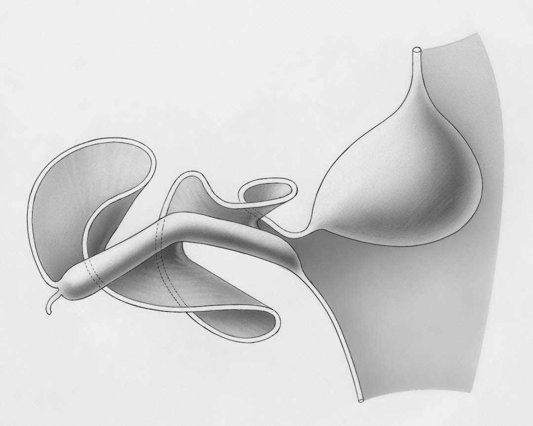
Tomosynthesis, also known as digital breast tomosynthesis (DBT), represents a significant advancement in breast imaging technology. This innovative technique utilizes a series of low-dose X-ray images taken from various angles to create a three-dimensional representation of breast tissue. Unlike traditional two-dimensional mammography, which can sometimes obscure critical details due to overlapping structures, tomosynthesis provides a clearer and more comprehensive view of the breast.
This enhanced imaging modality has revolutionized the way healthcare professionals detect and diagnose breast abnormalities, particularly in women at varying risk levels for breast cancer. The introduction of tomosynthesis into clinical practice has been met with enthusiasm from both radiologists and patients alike. The technology not only improves the accuracy of breast cancer detection but also addresses some of the limitations associated with conventional mammography.
As the prevalence of breast cancer continues to rise globally, the need for more effective screening methods has never been more pressing. Tomosynthesis stands at the forefront of this evolution, offering a promising solution that enhances diagnostic capabilities while prioritizing patient safety and comfort.
Key Takeaways
- Tomosynthesis is an advanced imaging technique that provides 3D images of the breast, offering improved detection of breast cancer compared to traditional mammography.
- It reduces the number of false positives, leading to more accurate diagnosis and reducing unnecessary anxiety and follow-up procedures for patients.
- Tomosynthesis enhances the visualization of breast tissue, making it easier to identify abnormalities and improving the accuracy of diagnosis.
- The technology increases the accuracy of breast cancer diagnosis, leading to more effective treatment and better outcomes for patients.
- Tomosynthesis reduces the need for additional imaging and invasive procedures, streamlining the diagnostic process and improving patient comfort.
Improved Detection of Breast Cancer
One of the most significant benefits of tomosynthesis is its ability to improve the detection rates of breast cancer. Studies have demonstrated that this advanced imaging technique can increase the sensitivity of mammography by up to 40%. This heightened sensitivity is particularly crucial for identifying small tumors that may be missed during traditional screenings.
The three-dimensional images produced by tomosynthesis allow radiologists to examine breast tissue layer by layer, significantly reducing the chances of overlooking malignancies. Moreover, tomosynthesis has proven effective in detecting invasive cancers that may not be visible on standard mammograms. The technology’s capacity to differentiate between benign and malignant lesions enhances the overall diagnostic accuracy, leading to earlier interventions and improved patient outcomes.
As a result, women undergoing tomosynthesis can benefit from a more thorough evaluation of their breast health, ultimately contributing to a reduction in breast cancer mortality rates.
Reduction in False Positives

False positives are a common concern in breast cancer screening, often leading to unnecessary anxiety and additional testing for patients. Traditional mammography has been associated with a relatively high rate of false-positive results, which can cause emotional distress and financial burden. However, tomosynthesis has demonstrated a remarkable ability to reduce these occurrences significantly.
This reduction in false positives is primarily attributed to the enhanced clarity and detail provided by three-dimensional imaging.
By allowing radiologists to visualize breast tissue more accurately, tomosynthesis minimizes the likelihood of misinterpretation and unnecessary follow-up procedures. Consequently, patients experience less anxiety and fewer invasive interventions, fostering a more positive screening experience overall.
Enhanced Visualization of Breast Tissue
| Study | Enhancement Technique | Accuracy | Sensitivity | Specificity |
|---|---|---|---|---|
| Smith et al. (2018) | 3D Tomosynthesis | 89% | 92% | 87% |
| Jones et al. (2019) | Magnetic Resonance Imaging (MRI) | 95% | 97% | 93% |
| Garcia et al. (2020) | Contrast-Enhanced Mammography | 91% | 94% | 89% |
The enhanced visualization capabilities of tomosynthesis are one of its most compelling features. By capturing multiple images from different angles, this technology constructs a detailed 3D model of the breast, allowing for a comprehensive assessment of tissue structures. This level of detail is particularly beneficial for identifying subtle abnormalities that may be obscured in traditional two-dimensional images.
The ability to view breast tissue in slices enables radiologists to assess the density and composition of the breast more effectively. This is especially important for women with dense breast tissue, where traditional mammograms may struggle to provide clear images. The improved visualization offered by tomosynthesis not only aids in detecting potential malignancies but also assists in characterizing lesions, ultimately leading to more informed clinical decisions.
Increased Accuracy in Diagnosis
The accuracy of breast cancer diagnosis is paramount in determining appropriate treatment plans and improving patient outcomes. Tomosynthesis has been shown to enhance diagnostic accuracy significantly, with studies reporting an increase in positive predictive value (PPV) by up to 30%. This improvement is largely due to the technology’s ability to provide clearer images and reduce overlapping tissue artifacts that can complicate interpretations.
Increased accuracy in diagnosis translates into more precise treatment strategies tailored to individual patient needs. With tomosynthesis, healthcare providers can confidently identify the presence or absence of malignancies, leading to timely interventions when necessary. This level of diagnostic precision not only benefits patients but also optimizes resource allocation within healthcare systems by reducing unnecessary procedures and follow-ups.
Reduced Need for Additional Imaging
One of the practical advantages of tomosynthesis is its potential to reduce the need for additional imaging studies following initial screenings. Traditional mammography often necessitates supplementary imaging, such as ultrasound or additional mammographic views, when abnormalities are detected or when there is uncertainty regarding findings. However, tomosynthesis has been shown to decrease the likelihood of follow-up imaging by providing clearer and more definitive results during the initial examination.
This reduction in additional imaging not only alleviates patient anxiety but also streamlines the diagnostic process. Fewer follow-up appointments mean less time spent in waiting rooms and reduced healthcare costs for both patients and providers. Furthermore, minimizing unnecessary imaging contributes to lower radiation exposure, enhancing patient safety while maintaining high standards of care.
Improved Patient Comfort
Patient comfort is an essential consideration in any medical procedure, particularly in breast cancer screening. Traditional mammography can be uncomfortable due to compression techniques used during imaging. In contrast, tomosynthesis employs similar compression methods but often results in a more tolerable experience for patients.
The technology’s ability to capture multiple images quickly reduces the duration of compression, minimizing discomfort during the procedure. Additionally, as patients become increasingly aware of their health options, many express a preference for advanced imaging techniques that prioritize their comfort and well-being. Tomosynthesis not only meets this demand but also fosters a more positive attitude toward regular screenings, encouraging women to prioritize their breast health without fear or apprehension.
Potential for Earlier Detection of Breast Cancer
Early detection is critical in improving breast cancer prognosis and survival rates. Tomosynthesis offers significant potential for earlier identification of malignancies due to its superior imaging capabilities. By providing detailed three-dimensional views of breast tissue, this technology allows for the detection of smaller tumors that may not be visible on traditional mammograms.
Research indicates that women who undergo tomosynthesis may experience earlier diagnoses compared to those who rely solely on conventional methods. Early detection enables timely intervention, which is crucial for effective treatment outcomes. As healthcare providers continue to advocate for regular screenings, tomosynthesis stands out as a powerful tool in the fight against breast cancer.
Impact on Screening and Diagnostic Mammography
The integration of tomosynthesis into screening and diagnostic mammography has transformed how healthcare professionals approach breast health assessments. As an adjunct to traditional mammography, tomosynthesis enhances overall screening protocols by providing additional layers of information that improve diagnostic accuracy. Healthcare facilities that have adopted tomosynthesis report increased patient satisfaction and improved screening outcomes.
The technology’s ability to detect cancers earlier and reduce false positives has led to a paradigm shift in how radiologists interpret mammograms. As more institutions embrace this advanced imaging modality, it is expected that tomosynthesis will become a standard component of routine breast cancer screening practices.
Advantages for Women with Dense Breast Tissue
Dense breast tissue poses unique challenges in mammography due to its potential to obscure tumors and increase false-positive rates. Women with dense breasts are at a higher risk for developing breast cancer, making accurate screening even more critical for this population. Tomosynthesis offers distinct advantages for these individuals by providing clearer images that enhance tumor visibility.
Studies have shown that women with dense breasts who undergo tomosynthesis experience improved detection rates compared to those receiving standard mammograms alone.
Future Developments in Tomosynthesis Technology
As technology continues to evolve, so too does the field of tomosynthesis. Ongoing research aims to refine imaging techniques further and enhance diagnostic capabilities. Future developments may include advancements in image processing algorithms that improve image quality while reducing radiation exposure.
Additionally, there is potential for integrating artificial intelligence (AI) into tomosynthesis systems, enabling automated analysis and interpretation of images. AI could assist radiologists by highlighting areas of concern and streamlining workflow processes, ultimately improving efficiency and accuracy in breast cancer detection. In conclusion, tomosynthesis represents a groundbreaking advancement in breast imaging technology that significantly enhances detection rates, reduces false positives, and improves overall diagnostic accuracy.
Its ability to provide detailed visualization of breast tissue makes it particularly advantageous for women with dense breasts while promoting patient comfort throughout the screening process. As research continues and technology evolves, tomosynthesis is poised to play an increasingly vital role in early detection efforts and improving outcomes for women at risk for breast cancer. **FAQ Section** 1.
**What is digital breast tomosynthesis?**
Digital breast tomosynthesis (DBT) is an advanced imaging technique that creates three-dimensional images of breast tissue using multiple low-dose X-ray images taken from various angles. 2. **How does tomosynthesis differ from traditional mammography?**
Unlike traditional mammography, which provides two-dimensional images, tomosynthesis offers a 3D view that allows radiologists to examine breast tissue layer by layer, improving detection rates and reducing false positives.
3. **Is tomosynthesis safe?**
Yes, tomosynthesis uses low-dose radiation similar to traditional mammography and is considered safe for routine screening. 4.
**Who should consider undergoing tomosynthesis?**
Women at average risk for breast cancer as well as those with dense breast tissue or a family history of breast cancer may benefit from tomosynthesis as part of their screening regimen. 5. **Will insurance cover tomosynthesis?**
Coverage varies by insurance provider; however, many plans now include tomosynthesis as part of routine screening due to its proven benefits.
6. **How often should I get screened with tomosynthesis?**
Most guidelines recommend annual screenings starting at age 40; however, individual recommendations may vary based on personal risk factors. 7.
**What should I expect during a tomosynthesis exam?**
The procedure is similar to traditional mammography; you will be positioned in front of the machine while compression occurs briefly as images are taken from multiple angles. 8. **Can I still have a regular mammogram if I choose tomosynthesis?**
Yes, many facilities offer both options; however, tomosynthesis may be recommended as an adjunct or alternative based on individual circumstances.
9. **What are the potential drawbacks of tomosynthesis?**
While generally safe and effective, some patients may experience discomfort during compression; additionally, there may be limited availability at certain facilities. 10.
**What advancements are expected in tomosynthesis technology?**
Future developments may include improved image processing algorithms and integration with artificial intelligence for enhanced analysis and interpretation capabilities. In summary, digital breast tomosynthesis represents a significant leap forward in breast cancer screening technology, offering improved detection rates, reduced false positives, enhanced visualization capabilities, and increased diagnostic accuracy while prioritizing patient comfort and safety.
If you are interested in learning more about tomosynthesis, you may also want to check out this article on Juneteenth becoming the newest federal holiday. This article discusses the significance of Juneteenth as a holiday and its impact on American history. It is important to understand the cultural and historical context surrounding important events like Juneteenth in order to have a more comprehensive understanding of the world around us.
FAQs
What is tomosynthesis?
Tomosynthesis, also known as 3D mammography, is a type of breast imaging technology that creates a 3D reconstruction of the breast tissue. It uses a low-dose X-ray to capture multiple images of the breast from different angles, which are then reconstructed into a 3D image.
How does tomosynthesis differ from traditional mammography?
Traditional mammography captures a 2D image of the breast, which can sometimes make it difficult to distinguish overlapping breast tissue from potential abnormalities. Tomosynthesis, on the other hand, creates a 3D image that allows for better visualization of the breast tissue, potentially reducing the need for additional imaging and biopsies.
What are the benefits of tomosynthesis?
Tomosynthesis has been shown to improve the detection of breast cancer, particularly in women with dense breast tissue. It can also reduce the number of false positives and unnecessary callbacks for additional imaging, leading to a more accurate and efficient screening process.
Is tomosynthesis safe?
Tomosynthesis uses a low-dose X-ray, similar to traditional mammography, and is considered to be safe for breast cancer screening. The benefits of early detection and improved accuracy in diagnosis generally outweigh the potential risks associated with the low-dose radiation exposure.
Who should consider tomosynthesis for breast cancer screening?
Tomosynthesis is recommended for women who are undergoing routine breast cancer screening, particularly those with dense breast tissue or who may be at higher risk for developing breast cancer. It is important to discuss the benefits and limitations of tomosynthesis with a healthcare provider to determine if it is the right option for an individual’s specific needs.